")
Analytical services

Host cell protein analysis
The presence of HCPs and added residuals - such as enzymes - may impact the safety and efficiency of your product. We can help you identify and quantify individual process-related impurities in the downstream process. This gives you a detailed risk assessment and enables efficient elimination of these proteins.

Product characterization
Combing the product characterization with an analysis of the process-related impurities gives a more thorough understanding of your molecule. This enables optimization of the process design to ensure the drug product achieves the required safety, purity, and potency attributes.
Biologic drugs
Enables characterization of recombinant
proteins and mAbs
Advanced therapies
Helps you understand impurities in complex products like cell, gene, and bacteriophage treatments
Vaccines
Use for analysis of live attenuated or inactivated
virus and VLP-based products
The Alphalyse approach
Get personal advice
Our skilled team of mass spectrometry experts offer exceptional advice and support. By taking time to understand your needs we'll assemble a customized service package that's focused on your goals. Think us of as an extension of your team: working with you, not just for you.


Access extensive knowledge
Thanks to our ever-growing range of client projects our protein analysis lab is constantly advancing and refining our techniques across expression systems and molecules. This means we can offer you detailed knowledge of essential product attributes and process-related residuals without you needing to invest in time-consuming training and method development.
Benefit from trusted LC-MS technology
Successful downstream optimization requires top-of-the-line instrumentation technology. Using our quantitative mass spectrometry assays you can progress your development process faster while saving on expensive equipment.

The Alphalyse approach
Get personal advice
Our skilled team of mass spectrometry experts offer exceptional advice and support. By taking time to understand your needs we'll assemble a customized service package that's focused on your goals. Think us of as an extension of your team: working with you, not just for you.
Access extensive knowledge
Thanks to our ever-growing range of client projects we're constantly advancing and refining our techniques across expression systems and molecules. This means we can offer you detailed knowledge of essential product attributes and process-related residuals without you needing to invest in time-consuming training and method development.
Benefit from trusted LC-MS technology
Successful downstream optimization requires top-of-the-line instrumentation technology. Using our quantitative mass spectrometry assays you can progress your development process faster while saving on expensive equipment.
You are in good company
Our clients include biotech enterprises, CMOs, and pharmaceutical companies in Europe, USA, and Canada.
The Alphalyse lab provides MS-based host cell protein analysis under GMP conditions which is approved for use as a release assay.

GMP
HCP service

year's experience

MS-based HCP projects

satisfied clients

reports delivered
You are in good company
Our clients include biotech enterprises, CMOs, and pharmaceutical companies in Europe, USA, and Canada.
The Alphalyse lab provides MS-based host cell protein analysis under GMP conditions which is approved for use as a release assay.
GMP
HCP service
year's experience
MS-based HCP projects
satisfied clients
reports delivered
What clients say

Thore Schmedt, Associate Director
AiCuris Anti-infective Cures AG, Germany

Max Kristiansen, MSc, Special Consultant Assay Development
Statens Serum Institut (SSI), Denmark

Kristiina Hyvärinen, Director QC, viral products
Targovax ASA, Finland
Torben Lund-Hansen, PhD, SVP
Head of Technical Operations
Y-mAbs Therapeutics Inc., USA

Lars Skriver
Senior Science Officer
SAVARA Aps, Denmark

Scott Kachlany, founder
Actinobac Biomed Inc.
Head of CMC, C> Division
GTP Bioways, France
What's new?
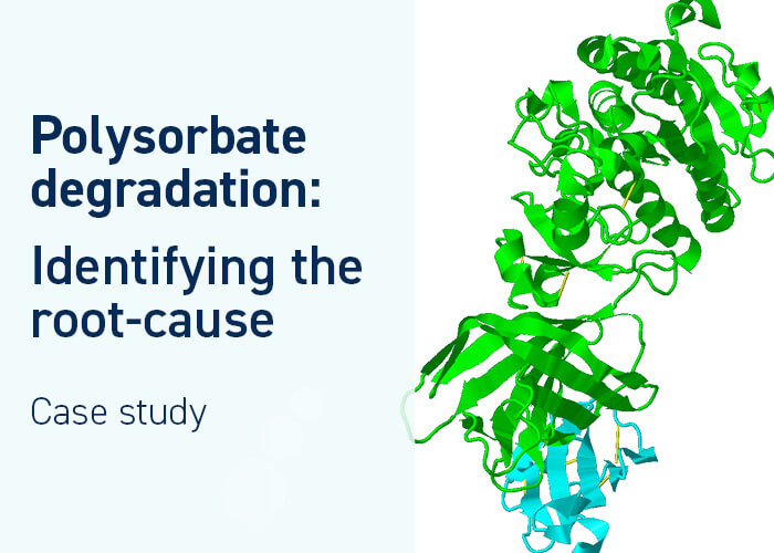
Video: What is causing polysorbate degradation?

Webinars: Genmab’s Holistic HCP Control Strategy for mAbs
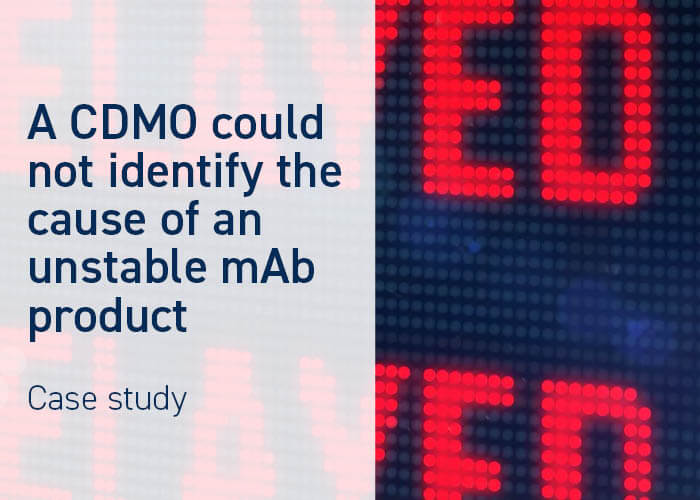
Video: Can your CDMO tackle Host Cell Proteins?

Blog: Why results obtained by new ELISA kit differ from the original

Exclusive Q&A: Insights from a leading CMC executive

Webinar: ELISA reagent characterization using LC-MS

Video: Rejected by the FDA → 2D gel Western blot was not enough

Insights from the BEBPA HCP Conference 2023
Webinar: LC-MS HCP assay validation and GMP release testing
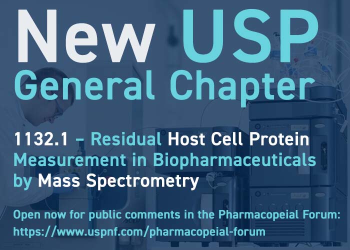
BREAKING: New USP general chapter on HCP analysis by MS

Selecting the best HCP-ELISA kit out of five
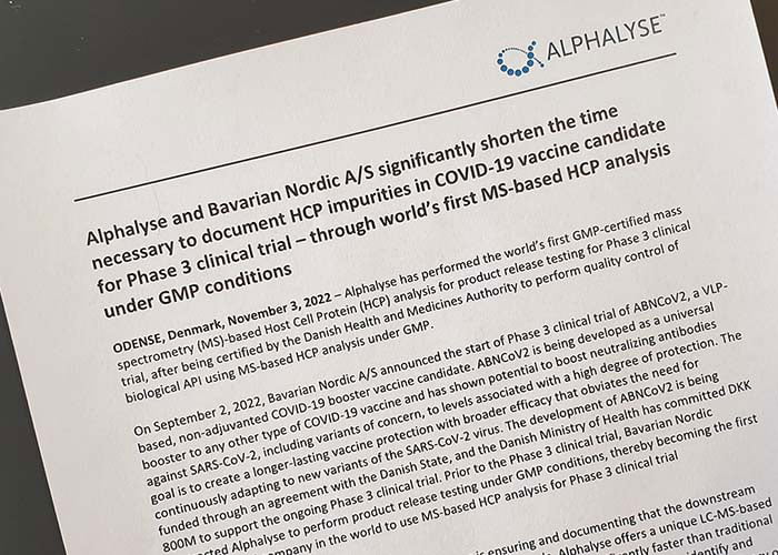
1st MS-based HCP analysis release test applied to COVID-19 candidate

Webinar: Analyzing vaccine purity – without an HCP ELISA

HCP-ELISA coverage analysis without a null cell line
Comparing viral protein quantities in AAV batches and DS

Stability study – an important part of biologics license application

Antibody characterization for cancer drug process development
Talk to us
Whatever protein-related challenge or question you may have, we would love to help. Our experts can help you decide on the best analytical approach for your project by email or online meeting - providing advice without obligation.